ឯកសារ:Diatoms.png

ទំហំរបស់ការមើលមុននេះ: ៧៣៤ × ៦០០ ភីកសែ។ ភាពម៉ត់ផ្សេងទៀត៖ ២៩៤ × ២៤០ ភីកសែ | ៥៨៨ × ៤៨០ ភីកសែ | ៩៤០ × ៧៦៨ ភីកសែ | ១២៥៣ × ១០២៤ ភីកសែ | ១៤០០ × ១១៤៤ ភីកសែ។
រូបភាពដើម (១៤០០ × ១១៤៤ ភីកសែល ទំហំឯកសារ៖ ៩៥១គីឡូបៃ ប្រភេទ MIME៖ image/png)
| ការពិពណ៌នា |
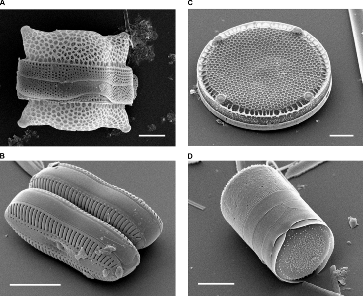
Scanning Electron Micrographs of Diatoms. (A) Biddulphia reticulata. The whole shell or frustule of a centric diatom showing valves and girdle bands (size bar = 10 micrometres). (B) Diploneis sp. This picture shows two whole pennate diatom frustules in which raphes or slits, valves, and girdle bands can be seen (size bar = 10 micrometres). (C) Eupodiscus radiatus. View of a single valve of a centric diatom (size bar = 20 micrometres) (D) Melosira varians. The frustule of a centric diatom, showing both valves and some girdle bands (size bar = 10 micrometres). |
||
| កាលបរិច្ឆេទ | Published: October 12, 2004 | ||
| ប្រភព | Bradbury J: Nature's Nanotechnologists: Unveiling the Secrets of Diatoms. PLoS Biol 2/10/2004: e306. doi:10.1371/journal.pbio.0020306 | ||
| អ្នកនិពន្ធ | Images courtesy of Mary Ann Tiffany, San Diego State University. | ||
| ការអនុញ្ញាត (ប្រើឯកសារនេះឡើងវិញ) |
|
ប្រវត្តិឯកសារ
ចុចលើម៉ោងនិងកាលបរិច្ឆេទដើម្បីមើលឯកសារដែលបានផ្ទុកឡើងនៅពេលនោះ។
| ម៉ោងនិងកាលបរិច្ឆេទ | កូនរូបភាព | វិមាត្រ | អ្នកប្រើប្រាស់ | យោបល់ | |
|---|---|---|---|---|---|
| បច្ចុប្បន្ន | ម៉ោង១៨:១០ ថ្ងៃព្រហស្បតិ៍ ទី១៦ ខែវិច្ឆិកា ឆ្នាំ២០០៦ | | ១៤០០ × ១១៤៤ (៩៥១គីឡូបៃ) | Ayacop | {{Information |Description='''Scanning Electron Micrographs of Diatoms.''' (A) ''Biddulphia reticulata''. The whole shell or frustule of a centric diatom showing valves and girdle bands (size bar = 10 micrometres). (B) ''Diploneis sp.'' This picture shows |
បម្រើបម្រាស់ឯកសារ
ទំព័រ ខាងក្រោមប្រើប្រាស់ឯកសារនេះ ៖
បម្រើបម្រាស់ឯកសារជាសាកល
វីគីដទៃទៀតដូចខាងក្រោមនេះប្រើប្រាស់ឯកសារនេះ៖
- ការប្រើប្រាស់នៅក្នុង ar.wikipedia.org
- ការប្រើប្រាស់នៅក្នុង ast.wikipedia.org
- ការប្រើប្រាស់នៅក្នុង bs.wikipedia.org
- ការប្រើប្រាស់នៅក្នុង ca.wikipedia.org
- ការប្រើប្រាស់នៅក្នុង cs.wikipedia.org
- ការប្រើប្រាស់នៅក្នុង de.wikipedia.org
- ការប្រើប្រាស់នៅក្នុង en.wikipedia.org
- ការប្រើប្រាស់នៅក្នុង es.wikipedia.org
- ការប្រើប្រាស់នៅក្នុង fr.wikipedia.org
- ការប្រើប្រាស់នៅក្នុង fr.wiktionary.org
- ការប្រើប្រាស់នៅក្នុង gl.wikipedia.org
- ការប្រើប្រាស់នៅក្នុង he.wikipedia.org
- ការប្រើប្រាស់នៅក្នុង ja.wikipedia.org
- ការប្រើប្រាស់នៅក្នុង nn.wikipedia.org
- ការប្រើប្រាស់នៅក្នុង outreach.wikimedia.org
- ការប្រើប្រាស់នៅក្នុង pl.wikipedia.org
- ការប្រើប្រាស់នៅក្នុង pt.wikipedia.org
- ការប្រើប្រាស់នៅក្នុង sk.wikipedia.org
- ការប្រើប្រាស់នៅក្នុង test2.wikipedia.org
- ការប្រើប្រាស់នៅក្នុង zh.wikipedia.org